CASE SUMMARY:
This case is based in California on a patient who has a Vocal Cord Tumor, and the treatment he received. A Standard of Care opinion will be needed LATER.
FIRST, I need a short Expert Opinion that helps establish, What Procedure Was Performed on the patients Vocal Cord on 1/5/15.
IMPORTANT:
Standard of Care has NOTHING to do with this initial opinion I need. This page should clarify exactly what I am requesting but please keep this in mind.
Based on your review of images/video and related medical records,
What Procedure Was Most Likely Performed on 1/5/2015?
1) Biopsy or similar procedure (5-20%+ removed, Most of Tumor remains)
OR
2) Tumor Removal (95-100% of Tumor removed)
In most civil cases, the legal requirement for stating an Expert Opinion is only “more than 50% probability.”
Your review will consist of mostly imaging, rather than written medical records. Here is whats included on this page:
1) – Images and Medical Notes from Stroboscopy on 9/12/13
2) – Images, Medical Notes and video from Stroboscopy on 10/30/14
3) – Images, Medical Notes and video from Stroboscopy on 5/27/15
4) – Images, Medical Notes and video from Stroboscopy on 5/10/18
5) – Images and Medical Notes from Stroboscopy on 6/4/18.
6) – CT Scan Notes from 9/17/13 and 5/31/18
7) – 2 Pathology Reports
REDACTED INFO: Any info not related to the Scopes or Tumor has been redacted. The physician and patient info has been redacted as well. This info will be disclosed as needed for opinion.
OBSERVATION #1
Based on images Before Procedure, no noticeable change occurred during Over 1 year from 9/12/13 to 10/30/14. The 10/30/14 medical notes below state as much, “It is unchanged from previous.”
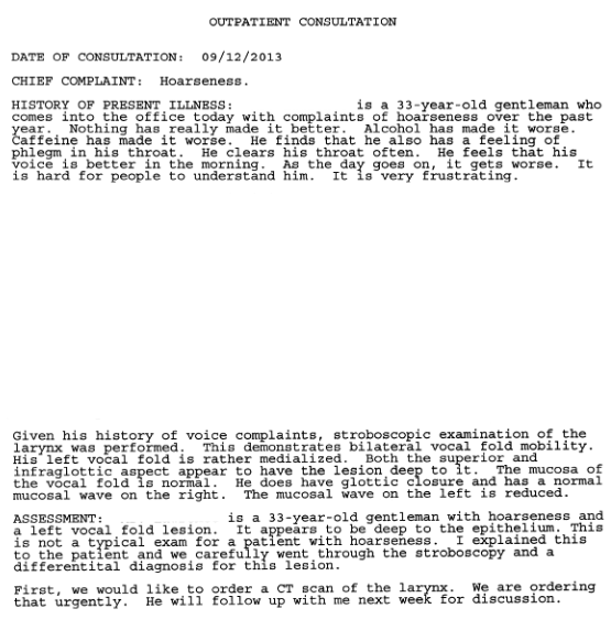
Images from a Scope on 9/12/2013, when Tumor was first diagnosed.
(If you click the image, a larger view will open in a new internet window)
")
 - Copy")
")
(If you click the Document image, it will open in a new internet window)
Scope Medical Records from 9/12/13
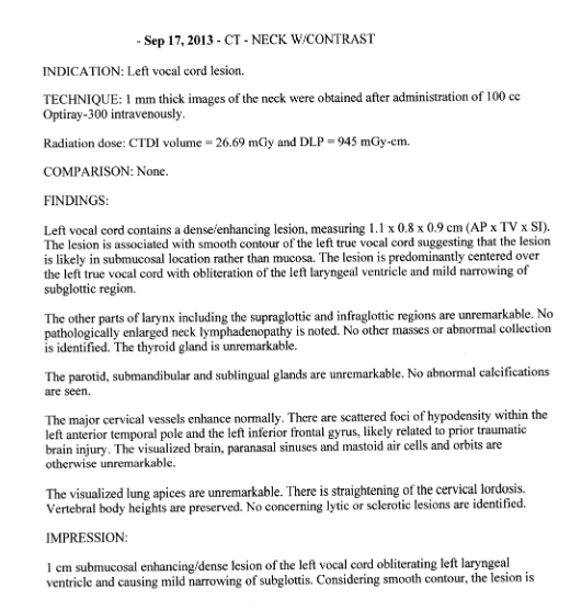
CT Scan Record from 9/17/13
Images from a Scope on 10/30/14, 2 months, 6 days Before Procedure
(If you click the image, a larger view will open in a new internet window)


(If you click the Document image, it will open in a new internet window)
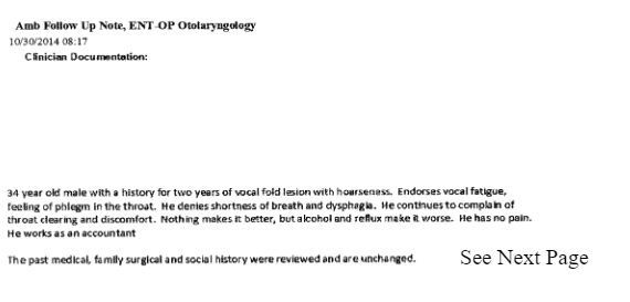
Scope Medical Records from 10/30/14
Recording of Scope 10/30/14 – click arrows on bottom right to increase to full screen.
The Procedure was performed on 1/5/2015

The full Procedure Report will not be provided as it should not have any influence.
OBSERVATION #2
Based on these images from 5/27/15 that are only 4 months and 22 days after Procedure, the Tumor still covers the left vocal cord and a good portion of patients airway. The size and appearance is not much different from the images above from before the 1/5/15 Procedure. If tumor was removed 95-100%, it appears to regrow very rapidly. (see below)
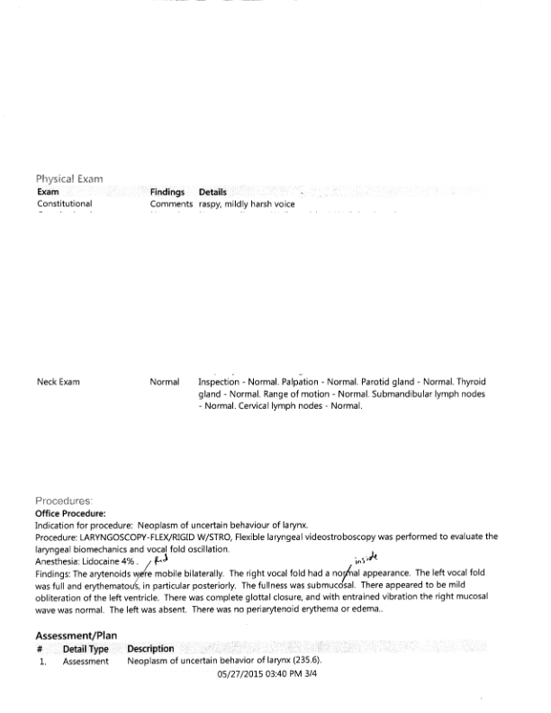
Images from a Scope on 5/27/15, 4 months, 22 days After Procedure
(If you click the image, a larger view will open in a new internet window)


(If you click the Document image, it will open in a new internet window)
Scope Medical Records from 5/27/15
Recording of Scope 5/27/15 – increase size by arrows on bottom right
Failed to load media. URL not valid. Please check WordPress Codex.
OBSERVATION #3
Based on images from 5/27/15 and 5/10/18, NO noticeable growth occurred during a 3 Year period after the Procedure. (see below)
Comparison of Scope images almost 3 years apart After Procedure
(If you click the image, a larger view will open in a new internet window)
THIS COLUMN – Scope images from 5/27/15, LESS than 5 months after Procedure
THIS COLUMN – Scope images from 5/10/18, Over 3 years after Procedure


(If you click the Document image, it will open in a new internet window)
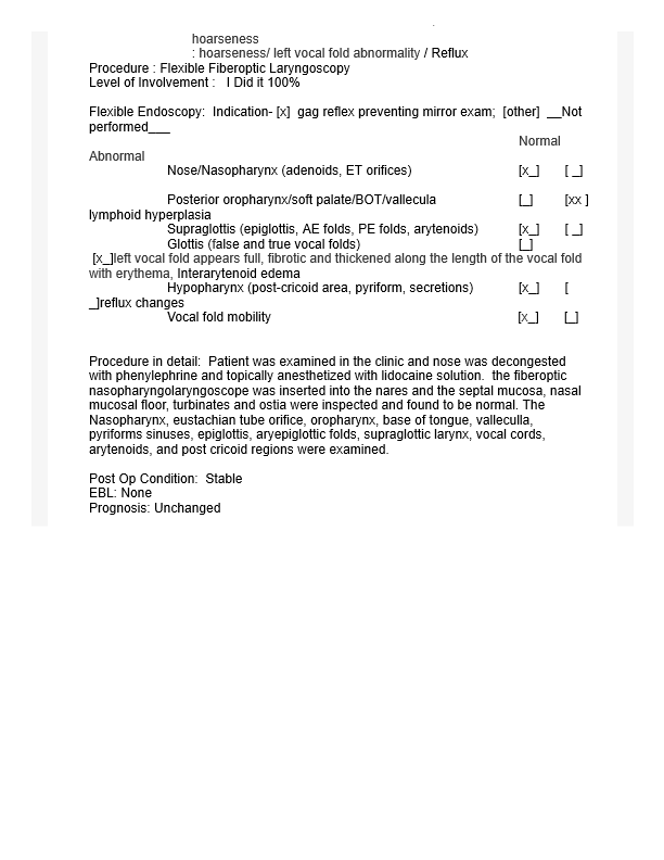
Scope Medical Records from 5/10/18
Recording of Scope 5/10/18 – click arrows on bottom right to increase to full screen.
OBSERVATION #4
From the first images post-procedure on 5/27/15 to 3 years later on 5/10/18 and 6/4/18, what looks like the incision area (bottom portion) does not grow back or fill in to the same thickness after over 3 years. The area removed is still easily noticeable, while regrowth in this area is NOT. It seems more consistent with a biopsy type Procedure and very little regrowth, if any.
(If you click the image, a larger view will open in a new internet window)
From Scope on 5/27/2015, Less than 5 months After Procedure
From Scope on 5/10/2018, 3 years 4 months after Procedure
From Scope on 5/10/2018, 3 years 4 months after Procedure
From Scope on 6/4/2018, 3 years 5 months after Procedure

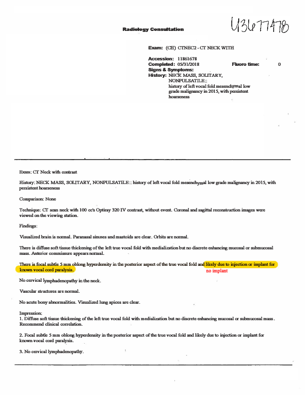
CT Scan from 5/31/18
Scope Medical Records from 5/10/18
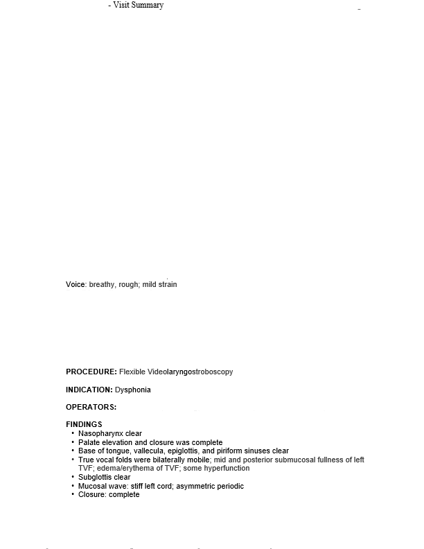
Scope Medical Records from 6/4/18
OBSERVATION #5
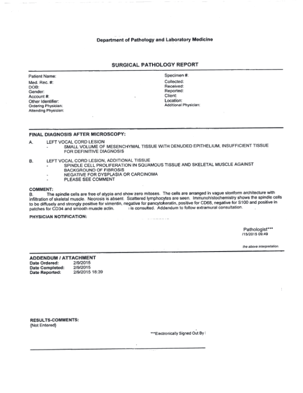
On the Pathology Reports, it states No/Negative/Zero for dysplasia, carcinoma, mitoses, necrosis, atypia, mitotic figures. Based on “degree of infiltration” ONLY, it was described as “low malignant potential” and diagnosed low grade malignancy by one pathologist. Additional opinions were sought and no definitive diagnosis was made. From review of Pathology alone, regrowth potential of this tumor appears very low and/or slow-growing. (see below)
(If you click on Document Image, a larger view will open in a new internet window)
Original Surgical Pathology Report
Emory Pathology Report